


 Foundational characteristics of cancer include proliferation, angiogenesis, migration, evasion of apoptosis, and cellular immortality. Find key markers for these cellular processes and antibodies to detect them.
 The SUMOplot™ Analysis Program predicts and scores sumoylation sites in your protein. SUMOylation is a post-translational modification involved in various cellular processes, such as nuclear-cytosolic transport, transcriptional regulation, apoptosis, protein stability, response to stress, and progression through the cell cycle.
 The Autophagy Receptor Motif Plotter predicts and scores autophagy receptor binding sites in your protein. Identifying proteins connected to this pathway is critical to understanding the role of autophagy in physiological as well as pathological processes such as development, differentiation, neurodegenerative diseases, stress, infection, and cancer.
HUMAN-SHB(Y268) Antibody
Purified Rabbit Polyclonal Antibody (Pab)
- SPECIFICATION
- CITATIONS
- PROTOCOLS
- BACKGROUND

Application
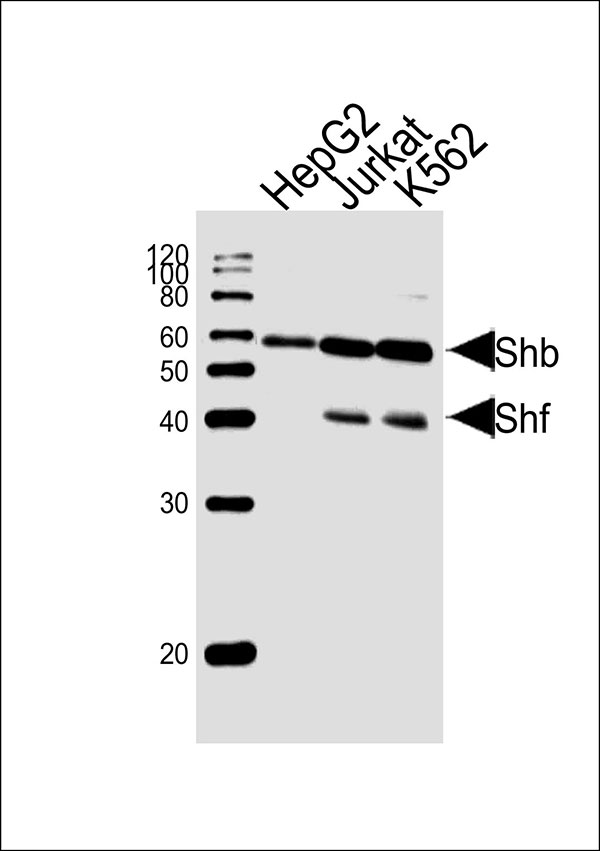
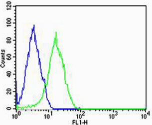
| WB, FC |
|---|---|
| Primary Accession | Q15464 |
| Reactivity | Human |
| Predicted | Mouse |
| Host | Rabbit |
| Clonality | polyclonal |
| Calculated MW | H=55,31;M=55 KDa |
| Isotype | Rabbit IgG |
| Antigen Source | HUMAN |
| Gene ID | 6461 |
|---|---|
| Antigen Region | 250-290 aa |
| Other Names | SH2 domain-containing adapter protein B, SHB |
| Dilution | WB~~1:1000 FC~~1:25 |
| Target/Specificity | This antibody is generated from a rabbit immunized with a KLH conjugated synthetic peptide between 250-290 amino acids from human. |
| Format | Purified polyclonal antibody supplied in PBS with 0.09% (W/V) sodium azide. This antibody is purified through a protein A column, followed by peptide affinity purification. |
| Storage | Maintain refrigerated at 2-8°C for up to 2 weeks. For long term storage store at -20°C in small aliquots to prevent freeze-thaw cycles. |
| Precautions | HUMAN-SHB(Y268) Antibody is for research use only and not for use in diagnostic or therapeutic procedures. |
| Name | SHB |
|---|---|
| Function | Adapter protein which regulates several signal transduction cascades by linking activated receptors to downstream signaling components. May play a role in angiogenesis by regulating FGFR1, VEGFR2 and PDGFR signaling. May also play a role in T-cell antigen receptor/TCR signaling, interleukin-2 signaling, apoptosis and neuronal cells differentiation by mediating basic-FGF and NGF-induced signaling cascades. May also regulate IRS1 and IRS2 signaling in insulin- producing cells. |
| Cellular Location | Cytoplasm. Cell membrane; Peripheral membrane protein; Cytoplasmic side. Note=Associates with membrane lipid rafts upon TCR stimulation |
| Tissue Location | Widely expressed.. |

Thousands of laboratories across the world have published research that depended on the performance of antibodies from Abcepta to advance their research. Check out links to articles that cite our products in major peer-reviewed journals, organized by research category.
info@abcepta.com, and receive a free "I Love Antibodies" mug.

Provided below are standard protocols that you may find useful for product applications.
Background
Adapter protein which regulates several signal transduction cascades by linking activated receptors to downstream signaling components. May play a role in angiogenesis by regulating FGFR1, VEGFR2 and PDGFR signaling. May also play a role in T-cell antigen receptor/TCR signaling, interleukin-2 signaling, apoptosis and neuronal cells differentiation by mediating basic- FGF and NGF-induced signaling cascades. May also regulate IRS1 and IRS2 signaling in insulin-producing cells.
References
Welsh M.,et al.Oncogene 9:19-27(1994).
Humphray S.J.,et al.Nature 429:369-374(2004).
Mural R.J.,et al.Submitted (SEP-2005) to the EMBL/GenBank/DDBJ databases.
Karlsson T.,et al.Oncogene 10:1475-1483(1995).
Karlsson T.,et al.Oncogene 13:955-961(1996).
If you have used an Abcepta product and would like to share how it has performed, please click on the "Submit Review" button and provide the requested information. Our staff will examine and post your review and contact you if needed.
If you have any additional inquiries please email technical services at tech@abcepta.com.
Ordering Information
Other Products
Shipping Information
